Each Icon below will take you to a page for the Respective ACLS EKG. These pages cover all of the cardiac arrhythmias that you will experience in the ACLS provider course. Rhythms from Ventricular Fibrillation to Complete Heart Block are covered. Examples of each ECG tracing are provided, and after each article is a short video that simulates the ACLS ECG on a defibrillator monitor.
You will also find a question and answer section below each rhythm video. This area covers the most common questions asked about each rhythm. Feel free to leave a comment or question as you review the content.
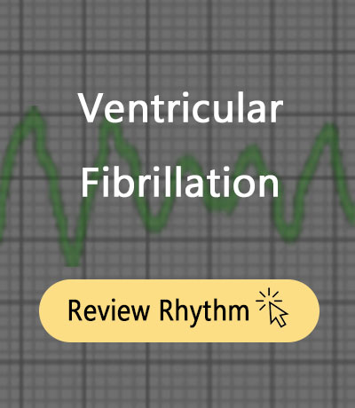
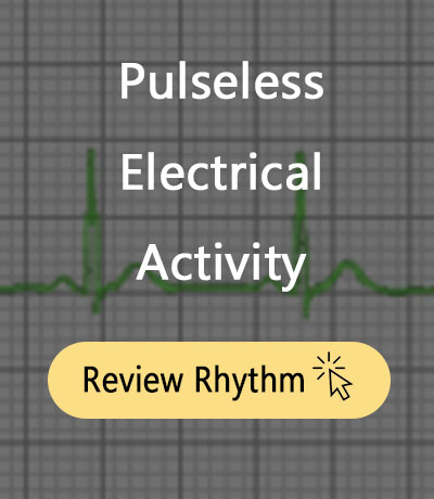
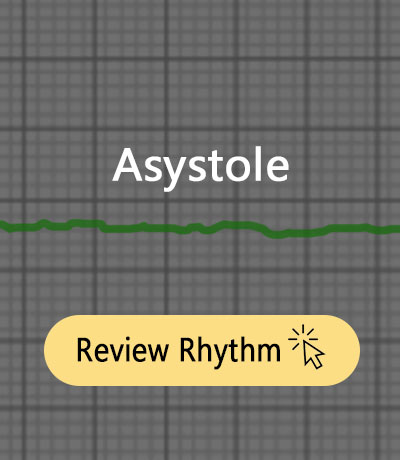
Pulseless Rhythms

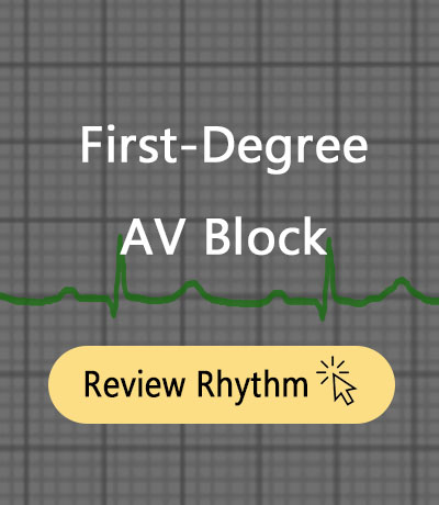
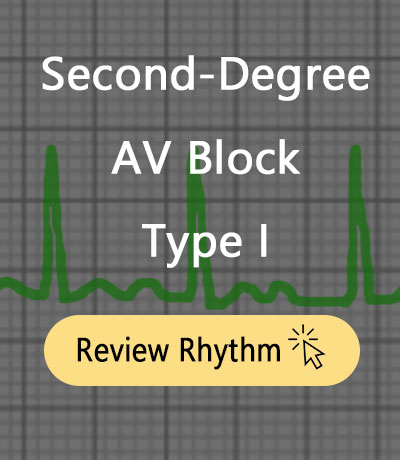
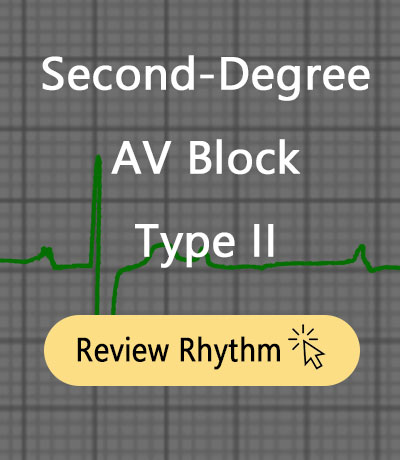
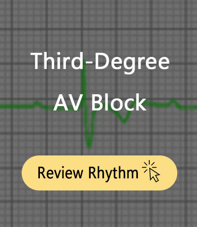
Bradyarrhythmias
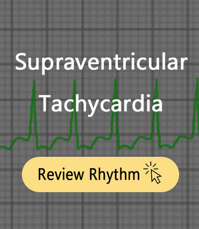
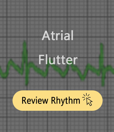
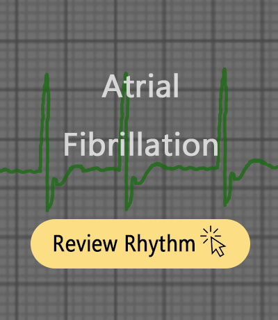
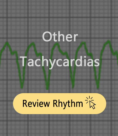
Tachyarrhythmias
Sheryl says
This was an excellent site. The rhythms were simple to review and understand. Thank you.
roseypumpkin says
Jeff,
Your site is wonderfully organized and easy to follow. It has helped me immensely!
Frances
Jeff with admin. says
Thanks for the encouraging feedback.
Kind regards,
Jeff
kayla says
agreed!!!!!!! great info
Susan Heidemann says
This site is so helpful I take my test the end of July, and I thank you so much for helping with the rhythms and the physiology behind them.
mrippeto says
Do we need to know the settings for synchronized cardioversions and defibrillation for the code simulations? I am having a hard time with this section any suggestions?
Maria
Jeff with admin. says
For ACLS, you should know the dose setting for both defibrillation and cardioversion of stable and unstable VT, SVT, and VF.
If you remember anything, remember the highlighted stuff.
For Defibrillation here is what you should remember: 120-200-300-360 (this is the standard increase in dosing)
For Cardioversion here is what you should remember:
(narrow complex regular) 50-100-200-300-360
(narrow irregular) 120-200-300-360
(wide complex regular) 100-200-300-360
(wide irregular) defibrillation same as VT/VF
The only difference is what is highlighted in red.
Kind regards,
Jeff
Robin says
So if out I the field and no aed, one would not know if it was a pulseless v tach, v fib or pea? Only by the monitor saying it is shockable that the differentiation happens?
Jeff with admin. says
That is correct. Until you have an AED or a defibrillator, there is no way to determine what kind of rhythm you are dealing with. In this case, you would initiate CPR with chest compression and rescue breaths. You would continue this while waiting for EMS or an AED.
Kind regards,
Jeff
Tina Williams says
I passed my ACLS test today! Thank you for this awesome site!
sara says
how long did you study and are there things that I might need to pay more attention to then others??? Thanks
Jeff with admin. says
I would recommend reading this.
Also use the acls-algorithms check-list to make sure that you cover everything on the site. You can download the checklist here.
Please let me know if you have any questions.
Kind regards,
Jeff
Debbie Pierce says
How can you tell the difference with coarse VT?
Jeff with admin. says
There is no such thing as coarse VT. Kind regards, Jeff
cpeer1 says
I love your website. I have learned so much! I am struggling with Monomorphic v-tach and polymorphic v-tach. Could you give me some suggestions to tell these apart.
Jeff with admin. says
Basically, with monomorphic VT all of the QRS complexes will look exactly the same. This is because all of the impulses are being generated from the same place in the ventricle.
With Polymorphic VT, the QRS complexes can appear different in size and position on the graph. This is because the impulse for each ventricular contraction can come from different locations with the ventricles.
This page may help also.
Kind regards,
Jeff
Chloe says
Love your site! Great study tool!
J Honea says
As a CNA I’m not qualified for ACLS, but the rhythms on your site are a great resource for ICU monitor tech training as well. Thank you for providing such a wonderful resource to the medical community.
mary bigsby says
Jeff, This site has been a God send! Would you consider adding more practice rhythms and interpretations for practice with treatment questions right after each strip as this part of the course always freaks me out!!! Thanks again and great job simplifying the course!
Jeff with admin. says
Yes Mary, Thanks for the encouragement. I will continue to add more practice rhythms and interpretations. Kind regards, Jeff
confusedperson says
I shadowed a cardiac floor for 2 days and i’m confused about a few things that the other nurses couldn’t really explain to me:
In some of the 5 lead rythmn strips, the complexts were facing downward, or, inverted. The nurse said the only reason for this was because of the lead we were looking at. I don’t understand why a lead would look like this.
And then at one point, a patient’s alarm was going off that they were in VTach. But the nurse said, “Oh, it’s not real VTach and cleared the alarms” she didn’t explain how she knew this by simply looking at the monitor. Please help me understand.
Jeff with admin. says
It sounds like you did not have a very good preceptor. He/She should have explained things to you as you shadowed them throughout the shift.
Your first question about “downward facing or inverted” QRS complexes: There are several reasons why you would see downward deflection on an ECG monitor. You were probably looking at Lead I, II, III, aVL, aVF, aVR. In a normal ECG, lead aVR is the only limb lead with a downwardly deflected QRS. The tracing of lead that is seen on the monitor or a rhythm strip is a representation of the movement of electricity through the heart. For a through understanding how an ECG works watch this.
As far at the nurse clearing the alarm when it alarmed “VTach.” It was most likely artifact that was being picked up from patient movement. When a person who is being monitored moves around more than a little this can significantly impact the way that the monitor is reads the patient’s heart. This can show up as wide and strange looking tracings which the monitor recognizes most of the time as ventricular tachycardia. Too bad the nursing did not take the time to tell you these things. Sorry for the bad experience. Don’t be afraid to ask for help from someone who will actually help.
Kind regards,
Jeff
Kyle says
I just wanted to say that this is an excellent site/resource for ACLS training. I’m trying to brush up before my course and this site has provided me with literally everything that I need. Thank you very much!
bhuvaneswari.k says
this site is very usefull for me
Maggie Hutchins says
Why is the ACLS test,course and instruction so hard to get done?
Jeff with admin. says
If this is your first time taking ACLS, it can be a little overwhelming. Use the checklist in the “download library” to make sure you cover everything. Also watch the video on the home page to see how to best utilize your time.
Kind regards,
Jeff